Cited by 827 — acetaldehyde; acetate; cytochrome P450 2E1 (CYP2E1); catalase; reactive oxygen species (ROS); blood alcohol concentration (BAC); liver; stomach; brain; fetal
184 KB – 10 Pages
PAGE – 1 ============
Overview: How I Metabolized b Samir Zakhari, Ph.D. SAMIR ZAKHARI, PH.D., is director, Division of Metabolism and Health Effects, National Institute on Alcohol Abuse and Alcoholism, Bethesda, Maryland. Alcohol is eliminated from the body by various metabolic mechanisms. The primary enzymes involved are aldehyde dehydrogenase (ALDH), alcohol dehydrogenase (ADH), cytochrome P450 (CYP2E1), and catalase. Variations in the genes for these enzymes have been found to influence alcohol consumption, alcohol-related tissue damage, and alcohol dependence. The consequences of alcohol metabolism include oxygen deficits (i.e., hypoxia) in the liver; interaction between alcohol metabolism byproducts and other cell components, resulting in the formation of harmful compounds (i.e., adducts); formation of highly reactive oxygen-containing molecules (i.e., reactive oxygen species [ROS]) that can damage other cell components; changes in the ratio of NADH to NAD+ (i.e., the cellÕs redox state); tissue damage; fetal damage; impairment of other metabolic processes; cancer; and medication interactions. Several issues related to alcohol metabolism require further research. KEY WORDS: acetaldehyde metabolism; alcohol dehydrogenase (ADH); aldehyde dehydrogenase (ALDH); acetaldehyde; acetate; cytochrome P450 2E1 (CYP2E1); catalase; reactive oxygen species (ROS); blood alcohol concentration (BAC); liver; stomach; brain; fetal alcohol effects; genetics and heredity; ethnic group; hypoxia The effects of alcohol (i.e., ethanol) on various tissues depend on its concentration in the blood (blood alcohol concentration [BAC]) over time. BAC is determined by how quickly alcohol is absorbed, distributed, metabolized, and excreted. After alcohol is swallowed, it is absorbed primar ily from the small intestine into the veins that collect blood from the stom- ach and bowels and from the portal vein, which leads to the liver. From there it is carried to the liver, where it is exposed to enzymes and metabolized. The rate of the rise of BA enced by how quickly alcohol is emp -tied from the stomach and the extent of metabolism during this first pass through the stomach and liver (i.e., first-pass metabolism [FPM]). BAC is influenced by environmen- tal factors (such as the rate of alcohol drinking, the presence of food in the stomach, and the type of alcoholic beverage) and genetic factors (variations in the principal alcohol-metabolizing enzymes alcohol dehydrogenase [ADH] and aldehyde dehydrogenase [ALDH2]). The alcohol elimination rate varies widely (i.e., three-fold) among individ- uals and is influenced by factors such as chronic alcohol consumption, diet, age, smoking, and time of day (Bennion and Li 1976; Kopun and Propping 1977). The consequent deleterious effects caused by equivalent amounts of alco- hol also vary among individuals. Even after moderate alcohol consumption, BAC can be considerable (0.046 to 0.092 gram-percent [g%]; in the 10- to 20-millimolar1 [mM] range). Alcoholreadily diffuses across membranes and distributes through all cells and tissues, and at these concentrations, it can acutelyaffect cell function by interacting with certain proteins and cell membranes. As explained in this article, alcohol metabolism also r tion of acetaldehyde, a highly r eactive and toxic byproduct that may contribute to tissue damage, the formation ofdamaging molecules known as reactive oxygen species (ROS), and a change in the reductionÐoxidation (or redox) state of liver cells. Chronic alcohol con- sumption and alcohol metabolism are strongly linked to several pathological consequences and tissue damage.Understanding the balance of alcoholÕs remov tially damaging metabolic byproducts, as well as how alcohol metabolism affects other metabolic pathways, is essentialfor appreciating both the short-term and long-term effects of the bodyÕs response to alcohol intake. Alcohol Metabolism Although the liver is the main organ responsible for metabolizing ingested alcohol, stomach (i.e., gastric) ADHhas been reported to contribute to FPM. The relativ ach and the liver to FPM, however, is controversial. Thus, whereas FPM is 1A millimole represents a concentration of 1/1,000 (onethousandth) molecular weight per liter (mol/L). Vol. 29, No. 4, 2006 245
PAGE – 2 ============
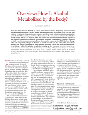
attributed pr ach (Lim et al. 1993; Baraona 2000), other previous studies (Lee et al. 2006) stress the role of the liver. Human ADH3, which is present in the liver and stomach, metabolizes alcohol poorly at physiological BACs (i.e., 0.23 g% BAC [or <50 mM]) in the liver but may play an important role in FPM in the stomach, because gastric alcohol concentrations can reach molar range during alcohol consumption (Baraona et al. 2001; Lee et al. 2003). However, Crabb (1997) pointed out the insuffi ciency of gastric ADH to account for FPM, so this remains unresolved. Alcohol also is metabolized in non liver (i.e., extrahepatic) tissues that do not contain ADH, such as the brain, by the enzymes cytochrome P450 and catalase (see below). I bolism is achieved by both oxidative pathways, which either add oxygen or remove hydrogen (through pathways involving ADH, cytochrome P450, and catalase enzymes), and nonoxidative pathways. Oxidative Pathways As shown in Figure 1, ADH, cytochrome P450 2E1 (CYP2E1), and catalase all contribute to oxidative metabolism of ethanol. ADH. The major pathway of oxidative metabolism of ethanol in the liver involves ADH (present in the fluid of the cell [i.e., cytosol]), an enzyme with many different variants (i.e., isozymes). Metabolism of ethanol with ADH pr duces acetaldehyde, a highly reactive and toxic bypr tribute to tissue damage and, possibly, the addictive process. As shown in Table 1, ADH constitutes a complex enzyme family, and, in humans, five classes have been categorized based on their kinetic and structural properties. A inated at a high rate because of the presence of enzyme systems with high activity levels ( Km),2 such as class II ADH, b3-ADH (encoded by ADH4 and ADH1B genes, respectively) and CYP2E1 (Bosron et al. 1993). This oxidation process involv diate carrier of electrons, nicotinamide adenine dinucleotide (NAD+), which is reduced by two electrons to form NADH. As a result, alcohol oxidation generates a highly reduced cytosolic environment in liv cytes). In other words, these reactions 2Km is a measurement used to describe the activity of an enzyme. It describes the concentration of the substance mal rate of reaction. Alcohol Research & Health 246 Figure 1 Oxidative pathways of alcohol metabolism. The enzymes alcohol dehydrogenase (ADH), cytochrome P450 2E1 (CYP2E1), and catalase all contribute to oxidative metabolism of alcohol. ADH, present in the fluid of the cell (i.e., cytosol), converts alcohol (i.e., ethanol) to acetaldehyde. This reaction involves an intermediate carrier of electrons, +nicotinamide adenine dinucleotide (NAD), which is reduced by two electrons to form NADH. Catalase, located in cell bodies called peroxisomes, requires hydrogen peroxide (H2O2) to oxidize alcohol. CYP2E1, present predominantly in the cellÕs microsomes, assumes an important role in metabolizing ethanol to acetaldehyde at elevated ethanol concentrations. Acetaldehyde is metabolized mainly by aldehyde dehydrogenase 2 (ALDH2) in the mitochondria to form acetate and NADH. ROS, reactive oxygen species.
PAGE - 3 ============
Alcohol Metabolism and the Body Table 1 Human Alcohol Dehydrogenase (ADH) Isozymes Class Gene Nomenclature Protein Km New Former mM Vmax min-1 Tissue I II III IV V ADH1A ADH1B*1 ADH1B*2 ADH1B*3 ADH1C*1 ADH1C*2 ADH4 ADH5 ADH7 ADH6 ADH1 ADH2*1 ADH2*2 ADH2*3 ADH3*1 ADH3*2 ADH4 ADH5 ADH7 ADH6 aaa b 1 b 2 b 3 g 1 g 2 p c s(m) 4.0 0.05 0.9 40.0 1.0 0.6 30.0 >1,000 30.0 ? 30 4 350 300 90 40 20 100 1,800 ? Liver Liver, Lung Liver, Stomach Liver, Cornea Most Tissues Stomach Liver, Stomach NOTE: The ADH1B and ADH1C genes have several variants with differing levels of enzymatic activity. Km is a measurement used to describe the activity of an enzyme. It describes the concentration of the substance upon which an enzyme acts that permits half the maximal rate of reaction. It is expressed in units of concentration. V max is a measure of how fast an enzyme can act. It is expressed in units of product formed per time. leave the liv ticularly vulnerable to damage from the byproducts of ethanol metabolism, such as free radicals and acetaldehy de. Cytochrome P450. The cytochrome P450 isozymes, including CYP2E1, 1A2, and 3A4, which are present pr dominantly in the microsomes, or vesi cles, of a network of membranes within the cell known as the endoplasmic r lum, also contribute to alcohol oxida tion in the liver. CYP2E1 is induced by chronic alcohol consumption and assumes an important r lizing ethanol to acetaldehyde at ele vated ethanol concentrations ( Km = 8 to 10 mM, compared with 0.2 to 2.0 mM for hepatic ADH). I dependent ethanol oxidation may occur in other tissues, such as the brain, where ADH activity is low. It also produces ROS, including hydroxyethyl, supero ide anion, and hydroxyl radicals, which increase the risk of tissue damage. Catalase. Another enzyme, catalase, located in cell bodies called peroxisomes, is capable of oxidizing ethanol in vitro in the presence of a hydrogen peroxide (H2O2)-generating system, such as the enzyme complex NADPH oxidase or the enzyme xanthine o dase. Quantitatively, however, this is considered a minor pathway of alcohol oxidation, except in the fasted state (Handler and Thurman 1990). Chronic alcohol consumption by rats has been shown to result in increased H 2O2 pro duction in pericentral regions of the liver and increased catalase activity (Misra et al. 1992). The role of CYP2E1 and catalase in alcohol metabolism in the brain are described in detail elsewhere (Zimatkin and Deitrich 1997). Products of Oxidative Metabolism of Alcohol Acetaldehyde and acetate, produced fr om the oxidative metabolism of alcohol, contribute to cell and tissue damage in various ways. Acetaldehyde. Acetaldehyde, produced by alcohol oxidation through any of the mechanisms outlined above, is rapidly metabolized to acetate, mainly b chondria), to form acetate and NADH. NADH then is oxidized by a series of chemical reactions in the mitochondria (i.e., the mitochondrial electr port chain, or respiratory chain). Acetaldehyde has the capacity to bind to proteins such as enzymes, microso mal proteins, and microtubules. It also forms adducts with the brain signaling chemical (i.e., neurotransmitter) dopamine to form salsolinol, which may contribute to alcohol dependence, and with DNA to form carcinogenic DNA adducts such as 1,N2-pr odeoxyguanosine. Formation of protein adducts in hepatocytes impairs protein secretion, which has been pro posed to play a role in enlargement of the liver (i.e., hepatomegaly). Acetate. Acetate, produced from the oxidation of acetaldehyde, is oxidized to carbon dioxide (CO 2). Most of the acetate resulting fr bolism escapes the liver to the blood and is eventually metabolized to CO 2 in heart, skeletal muscle, and brain cells. Acetate is not an inert product; it increases blood flow into the liver and depresses the central nerv tem, as well as affects various metabolic processes (Israel et al. 1994). Acetate also is metabolized to acetyl CoA, which is involved in lipid and cholesterol biosynthesis in the mitochondria of peripheral and brain tissues. I esized that upon chronic alcohol intake the brain starts using acetate rather than glucose as a source of energy . Nonoxidative Pathways The nonoxidative metabolism of alcohol is minimal, but its products may hav e pathological and diagnostic relevance. Alcohol is nonoxidatively metabolized by at least two pathways. One leads to the formation of molecules called fatty acid ethyl esters (FAEEs) from the r tion of alcohol with fatty acidsÐÐweak organic acids that play functional roles in human cells. The other nonoxidative pathway results in the formation of a taining phosphorus (i.e, phospholipid) known as phosphatidyl ethanol (see Figure 2). FAEEs are detectable in serum and other tissues after alcohol ingestion and persist long after alcohol is eliminated. The role of FAEEs in alcohol-induced tissue damage remains to be further evaluated. The second nonoxidative pathway requires the enzyme phospholipase D (PLD) (Laposata 1999), which breaks down phospholipids (primarily phos phatidylcholine) to generate phosphatidic acid (PA). This pathway is a criti cal component in cellular communication. Vol. 29, No. 4, 2006 247
PAGE – 4 ============
FAEE synthase Ethanol Fatty acid et hyl ester (FAEE) Phosphatidyl ethanol Tissue inju ry Interferes with PLD-dependent signaling? PLD Figure 2 Ethanol is nonoxidatively metabolized by two pathways. A reaction catalyzed by the enzyme fatty acid ethyl ester (FAEE) synthase leads to the formation of molecules known as FAEEs. A reaction with the lipid known as phosphatidyl ethanol. PLD has a high Km for ethanol, and the enzymatic reaction does occur pr dominantly at high circulating alcohol concentrations. The product of this reaction, phosphatidyl ethanol, is poorly metabolized and may accumulate to detectable levels follo wing chr sumption of large amounts of alcohol, but its effects on the cell remain to be established. Ho w ev er , the formation of phosphatidyl ethanol occurs at the expense of the normal function of PLD, namely to produce P A, r esulting in inhibited PA formation and disr uption of cell signaling. Oxidative and nono xidativ e pathways of alcohol metabolism are interr elated. Inhibition of ethanol o xidation b y com pounds that inhibit ADH, CYP2E1, and catalase results in an i n c r e a s e i n t h e nonoxidativ e metabolism o f a l c o h o l a n d increased pr oduction of F A E E s i n t h e liver and pancreas ( W erner e t a l . 2 0 0 2 ) . Genetic Aspects of Alcohol Metabolism V tribute significantly to clinical conditions observ ed after chr su mp ti on . These v ariations have been attributed to both genetic and envir mental factors, gender, drinking pattern, fasting or fed states, and chronic alcohol consumption. The following s ec ti on wi ll focus on the relev ant genetic f ac to rs . Genetic V ariation in ADH and ALDH Class I ADH and ALDH2 play a ce n t r a l r ole in alcohol metabolism. Variations in the genes encoding ADH and ALDH produce alcohol- and acetaldehyde-metabolizing enzymes that v ar y in activity . This genetic variability influences a personÕs su sc ep ti bi li ty to developing alcoholism and alcohol-related tissue damage. ADH. The ADH gene family encodes enzymes that metabolize v arious sub stances, including ethanol. The activity of these enzymes varies acr oss different organs (see Table 1). When ethanol is present, the metabolism of the other substances that ADH acts on may be inhibited, which may contribute to ethanol-induced tissue damage. As shown in T able 1, genetic v tion (i.e., polymorphism) occurs at the ADH1B and ADH1C gene locations (see Agarwal 2001), and these differ ent genes are associated with var ying levels of enzymatic activity. The ADH1B v ent frequencies in differ ent populati on s. For example, the ADH1B*1 form i s found predominantly in Caucasian and Black populations, wher eas ADH1B*2 frequency is higher in Chinese and Japanese populations and in 25 per cent of people with Je wish ancestry . ADH1C*1 and ADH1C*2 appear with roughly equal fr equency in Caucasian populations (Li 2000). People of J ewish descent carrying the ADH1B*2 allele show only marginally (<15 per cent) higher alcohol elimination rates compared with people with ADH1B*1 (Neumar k et al. 2001). Also, African Americans (Thomasson et al. 1995) and Nativ e Americans (W all et al. 1996) with the ADH1B*3 allele metabolize alcohol at a faster rate than those with ADH1B*1. ALDH. Sev eral isozymes of ALDH have been identified, but only the cytosolic ALDH1 and the mitochondrial AL DH 2 metaboliz e acetaldehy de. Ther morphism of the ALDH2 gene, r ing in allelic variants ALDH2*1 and ALDH2*2, which is virtually inactiv e. ALDH2*2 is present in about 50 per cent of the Taiwanese, H an Chinese, and Japanese populations (S hen et al. 1997) and shows vir hyde metabolizing activity in vitro . People who hav e one (i.e., heter gous) or especially two (i.e., homozygous) copies of the ALDH2*2 allele show incr eased acetaldehyde lev els after alcohol consumption (Luu et al. 1995; Wall et al. 1997) and ther efor ence negative physiological r esponses to alcohol. Because polymorphisms of ADH and ALDH2 play an important r ole in determining peak blood acetaldehyde levels and v oluntar tion (Quintanilla et al. 2005), they also in f dence. A fast ADH or a slow ALDH are expected to elevate acetaldehy de levels and thus r educe alcohol drinking. These polymorphisms and their signifi-Alcohol Research & Health 248
PAGE - 5 ============
Alcohol Metabolism and the Body cance are discussed in the article in this issue by Quertemont and Didone. ADH and ALDH isozyme activity also influences the prevalence of alcohol- induced tissue damage. Alcoholic cir rhosis is reduced more than 70 percent in populations carrying the ALDH2*2 allele (Chao et al. 1994; Nagata et al. 2002). In a review of studies, Yokoyama and Omori (2003) reported a positive correlation betw phisms for low-activity ADH and ALDH and esophageal and head and neck cancers. In another study (Hines et al. 2001), moderate drinkers who are homozygous for the slow-oxidizing ADH1C*2 allele, and therefore who are expected to drink at higher levels than those with the ADH1C*1 allele, showed a substantially decreased risk of heart attack (i.e., myocardial infarction). The authors (Hines et al. 2001, p. 549) only differentiated drinkers versus nondrinkers at one drink per day (ÒMen who con sumed at least one drink per day and were homozygous for the gamma2 allele had the greatest reduction in risk [relative risk 0.14]Ó). Interestingly, elevated acetaldehyde levels induced by ALDH inhibitors were shown to protect against alcohol- induced liver injury in experimental animals (Lindros et al. 1999) and to reduce the release of a signaling molecule (i.e., cytokine) called tumor necrosis factor alpha (TNF- a) from Kupffer cells (Nakamura et al. 2004). This finding is quite contradictory to the belief that acetaldehyde plays a r ole in liver damage. In a meta-analysis of most studies in the literature, Zintzaras and colleagues (2005) found that neither ADH nor ALDH alleles wer nificantly associated with liver cirrhosis. Genetic Variation in CYP2E1 Although several CYP2E1 polymorphisms have been identified, only a few studies were undertaken to determine the effect on alcohol metabolism and tissue damage. In one study (Ueno et al. 1996), the presence of the rare c2 hol metabolism in Japanese alcoholics but only at high BACs (0.25 g/dL). Raimondi and colleagues (2004) re ported that study participants with a polymorphism of CYP2E1 (CYP2E1 RsaI) were more likely than others to be lifetime abstainers at age 68 or older. Burim and colleagues (2004) found an association between having the m2/m2 CYP1A1 gene and alcoholic liv rhosis and the Val/Val GSTP1 thione S-transferase) gene and chronic pancreatitis. Consequences of Alcohol Metabolism The different pathways of ethanol metabolism described above have numerous detrimental consequences that contribute to the tissue damage and diseases seen in alcoholic patients. These consequences include oxygen deficits (i.e., hypoxia) in the liver; inter action between alcohol metabolism byproducts and other cell components, resulting in the formation of harmful compounds (i.e., adducts); formation of highly reactive oxygen-containing molecules (i.e., reactive oxygen species [ROS]) that can damage other cell components; and changes in the ratio of NADH to NAD+ (i.e., the cellÕs redox state [see Glossar sequences and the way they contribute to tissue damage and disease will be discussed in the following sections. Hypoxia As mentioned earlier, the main path way of alcohol metabolism, which involves ADH and ALDH, results in the generation of NADH. The NADH then is oxidized by a series of chemical reactions in the mitochondria (i.e., the mitochondrial electron transpor tem, or respiratory chain), eventually resulting in the transfer of electrons to molecular oxygen (O 2), which then binds protons (H +) to generate water (H2O). To have enough oxygen av able to accept the electr cytes must take up more oxygen than normal from the blood. Consistent with this assumption, studies have shown that ethanol metabolism tends to increase the hepatocytesÕ oxygen uptake from the blood ( Tsukamoto and Xi 1989). If the hepatocytes that are located close to the artery supplying oxygen-rich blood to the liver take up more than their normal share of o gen, however, not enough oxygen may be left in the blood to adequately supply other liver regions with oxygen. Indeed, strong evidence suggests that alcohol consumption results in significant hypoxia in those hepatocytes that are located close to the vein where the cleansed blood exits the liver (i.e., in the periv nous hepatocytes) (Arteel et al. 1996). The perivenous hepatocytes also are the first ones to sho age from chronic alcohol consumption (Ishak et al. 1991), indicating the potential harmful consequences of hypoxia induced by ethanol metabolism. In addition to directly increasing hepatocytesÕ oxygen use as described above, ethanol indirectly increases the cellsÕ oxygen use by activating Kupffer cells in the liver. When these cells become activated, they release various stimulatory molecules. One of these molecules is prostaglandin E2, which stimulates the metabolic activity of hepatocytesÑthat is, it induces them to break down and synthesize many essential molecules through a v ariety of chemical reactions that also require oxygen. As a result, alcohol-induced Kupffer cell activation also contributes to the onset of hypoxia. Adduct Formation Ethanol metabolism by ADH and CYP2E1 produce reactive molecules, such as acetaldehyde and ROS, that can interact with protein building blocks (i.e., amino acids) and other ble and unstable adducts (see Table 2). Acetaldehyde Adducts. Acetaldehyde interacts with certain amino acids in proteins (e.g., lysine, cysteine, and some of a group of amino acids called aromatic amino acids). However, not all amino acids in all proteins are equally likely to interact with acetaldehyde, and certain proteins seem to be par ularly susceptible to forming adducts with acetaldehyde. These include the following (Tuma and Casey 2003): Vol. 29, No. 4, 2006 249
PAGE - 6 ============
Table 2 Ethanol Metabolites and Adducts Generated During Ethanol Metabolism Source Ethanol metabolism Nonenzymatic lipid peroxidation of unsaturated fatty acids, breakdown of arachidonic acid in platelets Lipid peroxidation of long-chain polyunsaturated fatty acids Malondialdehyde-Acetaldehyde Hybrid adducts with malondialdehyde and Adduct (MAA) acetaldehyde Ethanol oxidation in the presence of iron ¥° Proteins found in the membranes surrounding the red blood cells (i.e., erythrocytes). ¥° Lipoproteins that consist of a pr tein and a fat component and which are associated with the risk of heart disease. ¥° Tubulin, a protein found in cell structures called microtubules that are essential for cell division and protein transport within cells. ¥° Hemoglobin, which is crucial for oxygen transport by the er ythrocytes. ¥° Albumin, which is a protein found in the blood. ¥° Collagen, the major pr nective tissue. ¥° Cytochrome enzymes, such as CYP2E1, which play a role in the metabolism of ethanol and many other substances. AcetaldehydeÐlysine adducts were detected in the plasma membrane of hepatocytes from alcohol-fed rats (Barry et al. 1987). These adducts can indirectly contribute to liver damage because the body recognizes them as ÒforeignÓ and therefore generates immune molecules (i.e., antibodies) against them. The presence of such antibodies has been demonstrated following chronic alcohol consumption (Israel et al. 1986). The antibodies bind to the adducts and induce the immune system to destro cytes containing these adducts. This process is known as immune-mediated hepatotoxicity or antibody-dependent cell-mediated cytotoxicity (ADCC). Antibodies directed against other acetaldehydeÐpr otein adducts also have been found in the blood of alcoholics (Lin et al. 1990; Worrall et al. 1990). Adducts formed by the interaction of acetaldehyde with erythrocyte membranes have been detected in the erythrocytes of alcohol abusers. These adducts may be associated with ethanol- induced macrocytosis, a condition characterized by unusually large numbers of enlarged erythrocytes in the blood (Niemela and Parkkila 2004). Macrocytosis is a marker for alcohol abuse. Finally, acetaldehyde can form adducts by interacting with compounds known as biogenic amines, 3 which include, among others, neur ters such as serotonin and dopamine. These adducts may hav cal effects on the nervous system. (For more information on adduct formation hyde, see the article by Quertemont and Didone, p. 258). ROS Formation. As mentioned earlier, ethanol metabolism by CYP2E1 and NADH oxidation by the electr port chain generate ROS that results in lipid peroxidation. This process results in the formation of compounds known as malondialdehy hydroxy-2-nonenal (HNE), both of which can form adducts with proteins (Worrall and Thiele 2001). I tion, acetaldehyde and MDA together can react with proteins to generate a stable MDAÐacetaldehydeÐprotein adduct (MAA) (Tuma et al. 1996; Tuma 2002). All of these adducts can induce immune responses (e.g., the formation of antibodies) (Tuma and Casey 2003). Moreover, MAA adducts can induce inflammatory processes in certain types of liver cells (i.e., stellate cells and endothelial cells) (Tuma 2002). These and other findings indicate a link between MDA, HNE, and MAA adducts and subsequent development of liver disease ( Tuma and Casey 2003). Formation of ROS and Decrease in Antioxidants ROS, including superoxide (O 2 ·Ð), hydrogen peroxide (H 2O2), hypochlorite ion (OClÐ), and hydroxyl ( ·OH) radicals, are naturally generated by many reactions in multiple regions of the cell. ROS act by ÒstealingÓ hydr gen atoms from other molecules, thereby converting those molecules into highly reactive fr natively , ROS can combine with stable molecules to form free radicals. Through both of these mechanisms, ROS play an important role in cancer dev ment (i.e., carcinogenesis), ather osis, diabetes, inflammation, aging, and other harmful processes. To prevent the damage these highly reactive compounds can cause, numerous defense systems have evolved in the body involving compounds called antioxidants, which can interact with ROS and conv ert them into harmless molecules. Under normal conditions, a balance between ROS and antioxidants exists in the cells. When this balance is disturbed and an excess of ROS is present, a state known as oxidative stress r esults. In most cells, the vast majority of ROS are generated in association with the mitochondrial electron transport system. In addition, ROS are produced by CYP2E1 and by activated Kupffer 3Biogenic amines are organic compounds formed during biochemical processes in plants and animals that carry a nitrogen atom as a central molecule. Alcohol Research & Health 250
PAGE - 8 ============
lized, and, second, electrons passing through the mitochondrial electron transport chain are ÒdivertedÓ into forming harmful R oxide (Hoek et al. 2002). Because ethanol metabolism by ADH and ALDH occurs primarily in the liver, any adverse effects associated with ethanol metabolism by these enzymes and associated ROS pr marily would affect that organ. In contrast, CYP2E1, which also oxidizes ethanol, particularly following chronic alcohol intake, is found in many tissues in addition to the liver, including the brain, heart, lungs, and certain white blood cells (i.e., neutrophils and macrophages). Accordingly, mediated ethanol oxidation would affect numerous tissues. Harmful effects asso ciated with CYP2E1-mediated ethanol metabolism primarily are related to the production of ROS, mainly superoxide and hydroxyl radicals. This ROS pr duction contributes to alcohol-induced damage to a variety of tissues not only by causing oxidative stress but also by enhancing apoptosis triggered by v ous stimuli. In the liver mediated ethanol metabolism generates oxidative str age and may thereby play an important role in alcohol-related development of liver cancer (Bradford et al. 2005). Effects on Fetal Development Oxidative stress plays an important role in ethanol-induced damage to the developing fetus (Cohen-Kerem and Koren 2003). Low levels of CYP2E1 are found in prenatal brain (Brezezinki derived ROS could play a role in the development of alcohol-related birth drome (FAS). M oreo ver, ROS pr duced during CYP2E1-mediated ethanol metabolism would likely be particularly harmful because the fetal brain shows only low levels of antio dant enzyme activity compared with adult brain (Henderson et al. 1999). Researchers have studied whether administration of antioxidants, such as N-acetyl cysteine, SAMe, folic acid, and vitamin C, could improv vival during fetal ethanol exposure; however, these studies have yielded mixed results. Nonoxidative metabolism of ethanol b cated in alcohol-related birth defects. As mentioned earlier, phospholipase D lular signal transduction processes, and the presence of ethanol interferes with bition of these signaling pr ing fetal development impairs the pro liferation of certain brain cells (i.e., astroglial cells) and may contribute to the reduced brain size (i.e., micr cephaly) found in most childr nosed with FAS (Costa et al. 2004). Impairment of Other Metabolic Processes Chronic ethanol consumption and alcohol metabolism also may influence various other metabolic pathways, thereb orders frequently found in alcoholics, such as fatty liver and excessive levels of demia), accumulation of lactic acid in the body fluids (i.e., lactic acidosis), excessive pr pounds known as ketones in the body (i.e., ketosis), and elevated levels of uric acid in the blood (i.e., hyperuricemia). The liver is most commonly affected by alcohol-induced damage. The first stage of liver damage following chr onic alcohol consumption is the appearance of fatty liver, which is followed by inflammation, apoptosis, fibrosis, and finally cirrhosis. The development of fatty liver is induced by the shift in the redox state of the hepatocytes that results from ethanol metabolism by ADH. This shift in the redox state favors the accumulation of fatty acids, rather than their oxidation. In addition to these metabolic effects, chronic ethanol consumption contributes to the development of fatty liver b ing the activities of several proteins that help regulate fatty acid synthesis and oxidation (Nagy 2004). O ciated with ethanol metabolism result from the fact that ADH and ALDH metabolize not only ethanol but also other compounds. For example, ADH and ALDH oxidize r min A1) to retinal and, subsequently, retinoic acid, which plays an important role in growth and differentiation. In the presence of ethanol, ADH and ALDH may be occupied with ethanol metabolism and retinol metabolism may be inhibited. These interactions may have serious implications for fetal development, stem cell differentiation, maintenance of differentiated tissue function, and the normal structure and function of stellate cells in the liver (Crabb et al. 2001). Chronic alcohol consumption also is associated with disturbances in the metabolism of sulfur-containing amino acids, leading to increased levels of the amino acids glutamate, aspartate, and homocysteine in alcoholic patients. These increases may have serious adverse effects. F teine increases and modulates cer tain nerve signaling processes, particularly during alcohol withdrawal, and increases in homocysteine levels may possibly contribute to the alcoholism-associated tissue shrinkage (i.e., atrophy) obser ved in brain tissue (Bleich et al. 2004). Cancer Risk and Medication Interactions Chronic alcohol consumption greatly enhances the risk of developing cancer of the esophagus and oral cavity (Seitz et al. 2004) and plays a major role in the development of liver cancer (Stickel et al. 2002). Several mechanisms have been identified that contribute to ethanol-associated tumor development, some of which are related to alcohol metabolism. F hyde generated during alcohol metabolism promotes cancer dev elopment, as does induction of CYP2E1 leading to ROS formation (Pıschl and Seitz 2004). Induction of CYP2E1 following heavy alcohol consumption also has other potentially harmful consequences: ¥° Enhanced alcohol metabolism by CYP2E1 contributes to alcoholicsÕ Alcohol Research & Health 252
184 KB – 10 Pages